In vivo imaging laboratory
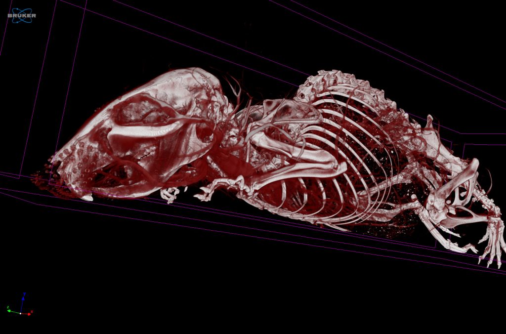
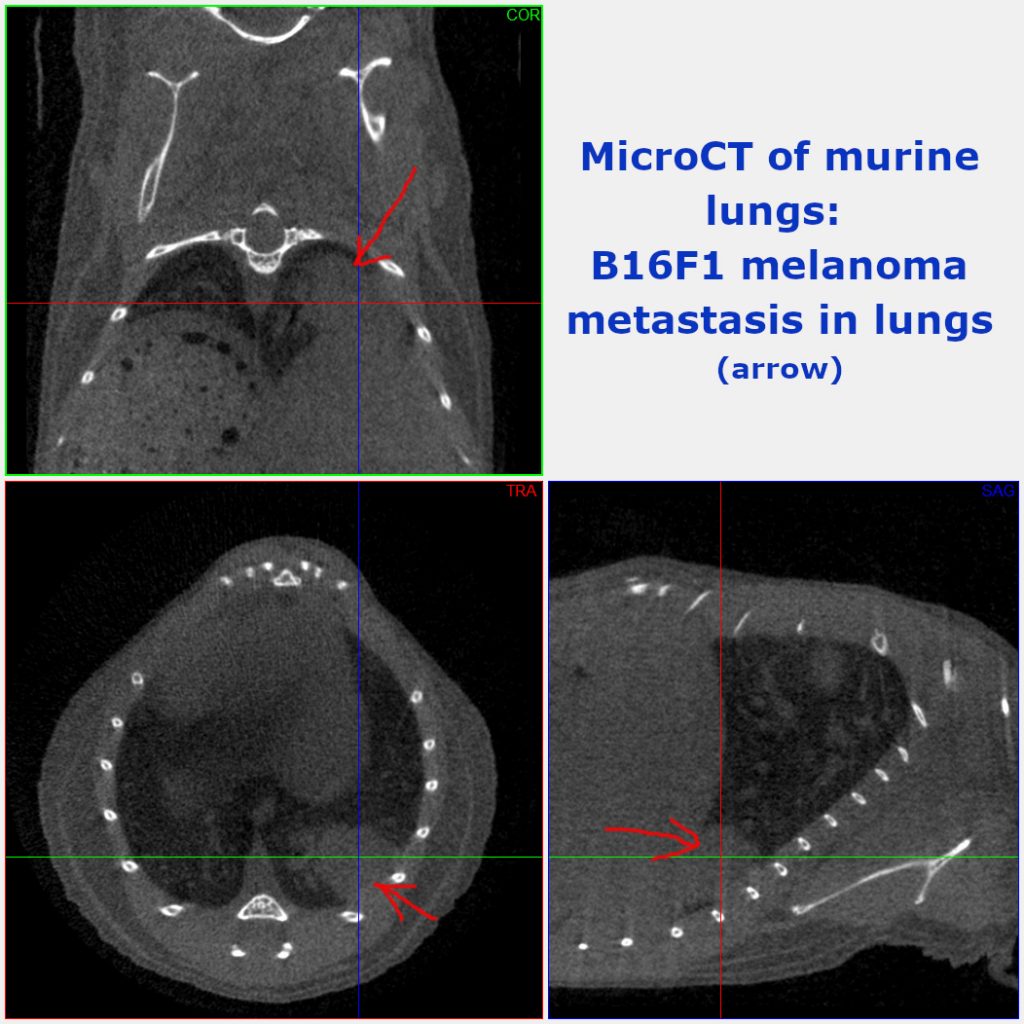
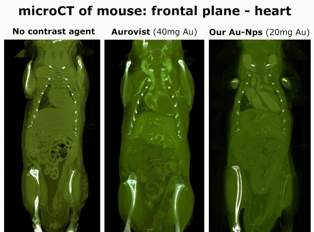
To observe and characterize patho-morphologic changes, distribution of active compounds and pathogens in organisms, we use noninvasive whole-body imaging techniques, including X-ray microtomography, in vivo fluorescence and bioluminescence. Small-animal whole-body in vivo imaging allows us to simultaneously monitor continuous biological processes in the same animal and can also reduce the number of animals for experimentation. Thus, it is in agreement with 3R principles. We use both mice and rat models in our lab. In addition, imaging modalities can serve for some ex-vivo and in-vitro studies.
Equipment
Current research topics
Head of the laboratory

Cooperation offer
We offer whole-body in-vivo/ ex-vivo imaging in rodent models – tracking and distribution of labeled active compounds or immune cells in organism cancer models
Our partners